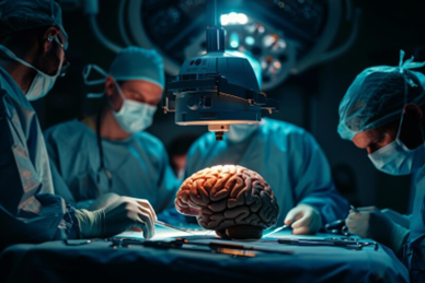
A craniotomy is a surgical procedure that involves making an opening in the skull to access the brain. This operation is performed by a neurosurgeon and is typically necessary for treating conditions like brain tumors, aneurysms, and traumatic brain injuries.
Types of Craniotomy
Craniotomies can be categorized based on the area of the skull being accessed:
Frontal Craniotomy: Through the frontal bone at the forehead.
Temporal Craniotomy: Through the temporal bone at the temples.
Parietal Craniotomy: Through the parietal bones on either side of the skull.
Occipital Craniotomy: Through the occipital bone at the back of the head.
Pterional Craniotomy: At the junction of the frontal, temporal, and parietal bones.
Additional specialized techniques include:
Keyhole Craniotomy: A minimally invasive approach using a small incision.
Stereotactic Craniotomy: Utilizing 3D imaging to locate the problem area.
Awake Craniotomy: The patient is conscious during the procedure to monitor critical brain functions.
Indications for Craniotomy
A craniotomy may be performed for several reasons, including:
- Removal of brain tumors or blood clots.
- Clipping of aneurysms to prevent rupture.
- Surgery for epilepsy or arteriovenous malformations.
- Ventricular shunting to reduce pressure from excess fluid.
Preoperative Preparations
Before undergoing a craniotomy, patients will typically have several assessments, including:
- Imaging Tests: CT or MRI scans to locate the issue.
- Blood Tests: To check hemoglobin levels and other vital markers.
- Other Tests: Urinalysis, ECG, and possibly a chest X-ray, especially for older patients.
Patients are usually required to fast the night before surgery and may be prescribed medications to manage seizures or swelling.
Procedure Overview
Craniotomy usually involves several steps:
Anesthesia: General anesthesia is administered, ensuring the patient is asleep and unaware during the procedure.
Incision: A scalp incision is made at the surgical site, with hair often shaved in the area.
Skull Opening: A piece of the skull (bone flap) is removed using a medical drill and saw.
Accessing the Brain: The dura mater, the brain’s outer membrane, is cut to access the brain for treatment.
Closure: After surgery, the bone flap is replaced, and the scalp is sutured closed.
Recovery Process
Post-surgery, patients are monitored in a recovery unit or intensive care. Typical recovery includes:
- Hospital stay of about one week.
- Ongoing monitoring of neurological function.
- Physical, occupational, or speech therapy to regain abilities.
At home, follow-up appointments are crucial for monitoring recovery, and lifestyle adjustments like a healthy diet, regular exercise, and avoiding smoking can help ensure long-term health.
Risks and Complications
Like any major surgery, craniotomy carries risks, such as infection, bleeding, and neurological issues. It is vital to report any severe headaches, seizures, or signs of infection to your doctor immediately.
In summary, a craniotomy is a significant but often necessary procedure for addressing serious brain conditions. Following medical advice and maintaining follow-up care is crucial for a successful recovery and improved quality of life.


